Introduction
Fixed Appliance treatment can significantly alter and improve facial appearance in addition to correcting irregularity of the teeth. Class I malocclusion is more prevalent than any type of malocclusion . The number of adults seeking orthodontic treatment has increased significantly.1, 2 Treatment alternatives of correction of a Bimaxillary Dentoalveolar protrusion are either Orthodontic camouflage by extraction of premolars or a Combined orthodontic-orthognathic surgical therapy. It eventually depends mainly upon the severity of the malocclusion 3, 4 and the amount of needed tooth movements. 3, 5 If the skeletal discrepancy 6 cannot be corrected by camouflage, any dental compensation may produce a reasonably good occlusion 7 but at the expense of compromised esthetics. For adult patients having severe orthodontic problems, surgery to realign the jaws or reposition dentoalveolar segments is the only possible treatment option left. One indication for surgery is a malocclusion so severe that it cannot be corrected by orthodontics alone. 8 Over the last few decades, there are increased number of adults who have become aware of orthodontic treatment and are demanding high quality treatment, in the shortest possible time with increased efficiency and reduced costs. Class I malocclusion patients frequently show a combinations of skeletal and dentoalveolar components. Many cephalometric pecularities have been reported in class I Bimax patients, such as a prognathic maxilla and mandible, proclined maxillary and mandibular incisors, an increased lower anterior face height and obtuse gonial angle. 9, 10 Prevalence of class I malocclusion in caucasians ranges from 0.8% to 4.0% and increases up to 12-13% in Chinese and Japanese population, while in American population class I malocclusion ranges from 3-4% of the population. 9, 10, 11 The correction of Bimaxillary prognathism is a procedure that dates back more than 10 decades. In 121849 Hullihen described a technique for the correction of such a deformity. Since that time refinements of technique and various methods have 13 been described. At the turn of the century Blair published several articles on this particular subject. Interest in the subject and in the various techniques used in its correction became widespread. After 14, 15 Blair, came reports from Kazanjian , Dingman, 16, 17 Reiter,” Caldwell and Letterman, Moose, and many others. This case presents the correction of a Bimaxillary dentoalveolar protrusion with a Class I malocclusion in a growing male patient with moderate crowding and severely proclined maxillary and mandibular anterior teeth merely simply by executing extraction of maxillary and mandibular 1st premolars. The Extraction protocol shown in this case is indicative of how a convex unesthetic facial profile can be converted into an Orthognathic pleasant profile by routine Fixed Orthodontic treatment with extraction of 4 premolars followed by retraction and closure of spaces.
Case Report
Extra-Oral Examination
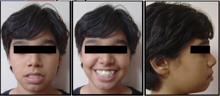
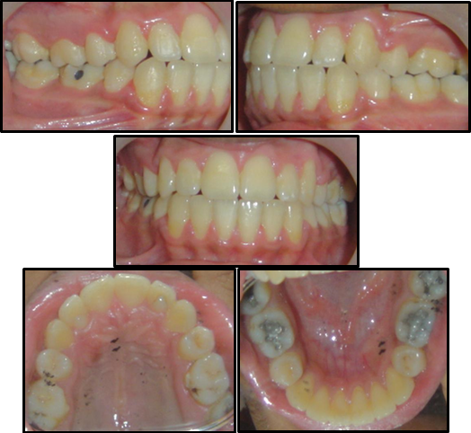
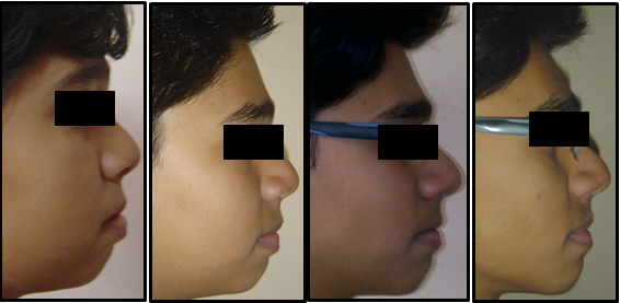
A 15 year old male patient presented with the chief complaint of forwardly placed upper and lower front teeth and excessive show of front teeth. On Extraoral examination, the patient had a convex facial profile, grossly symmetrical face on both sides with a retruded chin, potentially incompetent lips ,moderately deep mentolabial sulcus and an acute Nasolabial Angle , a Mesoprosopic facial form, Dolicocephalic head form, Average width of nose and mouth, minimal buccal corridor space, a consonant smile arc and slightly anterior divergence of face. The patient had no relevant prenatal, natal, postnatal history, history of habits or a family history. On Smiling, there was excessive show of maxillary anterior teeth. The patient had a toothy smile. On smiling the patient showed the presence of acrowded anterior dentition and an unaesthetic facial profile with a toothy smile.
Intra-Oral Examination
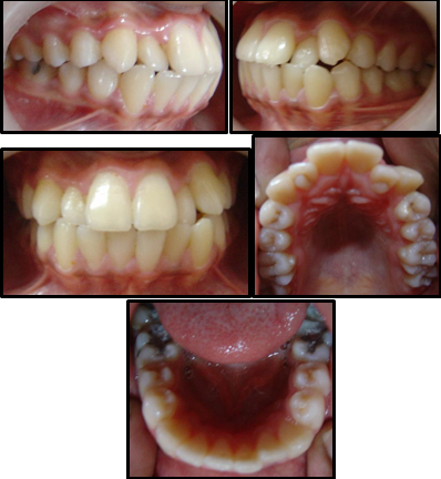
Intraoral examination on frontal view shows presence of an average overjet and overbite with the lower dental midline shifted slightly to the left of the patient y 1mm. On lateral view the patient shows the presence of Class I incisor relationship, a Class I Canine relationship bilaterally and a Class I molar relationship bilaterally. There was slight crowding in the upper arch with presence of instanding lateral incisors and severely protruded upper and lower anterior dentition. The upper and lower arch showed the presence of a U shaped arch form. Also the patient showed presence of a bilateral single tooth crossbite with the right and left lateral incisors. Also the right and left lateral incisors showed the presence of a Talons cusp on occlusal view
Table 1
Pre treatment cephalometric readings
Steiners analysis shows a prognathic maxilla and a prognathic mandible, Class I Skeletal pattern, a horizontal growth pattern, proclined maxillary and mandibular anteriors, forwardly placed maxillary and mandibular anteriors and protrusive upper and lower lips
Tweeds analysis shows a Horizontal growth pattern and proclined mandibular incisors
Wits appraisal shows AO and BO coinciding indicating Skeletal Class I pattern
Ricketts analysis shows a horizontal growth pattern ,average positioned condyles and proclined maxillary and mandibular anteriors
McNamara analysis shows a prognathic maxilla and mandible, a horizontal growth pattern, average lower anterior facial height, an acute Nasolabial Angle and proclined maxillary and mandibular incisors
Rakosi Jaraback analysis shows a horizontal growth pattern and proclined maxillary and mandibular incisors
Holdaway soft tissue analysis shows increased maxillary and mandibular sulcus depth and increased strain of lips along with a retruded chin position
Downs analysis shows a Class I Skeletal pattern, a horizontal growth pattern and proclined maxillary and mandibular anterior teeth
Diagnosis
This 15 year old male patient was diagnosed with a I malocclusion with a prognathic maxilla and mandible and a horizontal growth pattern, average overjet and overbite, proclined upper and lower incisors, crowding in the upper anterior region, a retruded chin, moderately deep mentolabial sulcus and potentially incompetant lips and a convex facial profile.
Treatment Objectives
To correct maxillary and mandibular prognathism
To correct proclined maxillary and mandibular anterior teeth
To correct the posterior divergence of face
To correct crowding in the maxillary anterior teeth
To correct the retruded chin position
To correct the decreased Nasolabial angle
To correct the dental midlines
To decrease the lip strain
To achieve a pleasing smile and a pleasing profile
Treatment Plan
Extraction of 14, 24, 34 and 44
Fixed appliance Therapy with MBT 0 022 inch bracket slot
Initial leveling and alignment with 0.012”, 0.014”, 0.016”, 0.018”, 0.020” Niti archwires following sequence A of MBT
Retraction and closure of spaces by use of 0.019” x 0.025” rectangular NiTi followed by 0.019” x 0.025” rectangular stainless steel wires
Final finishing and detailing with 0 014” round stainless steel wires
Retention by means of Beggs Wrap-around retainers along with lingual bonded retainers in the upper and lower arch.
Treatment Progress
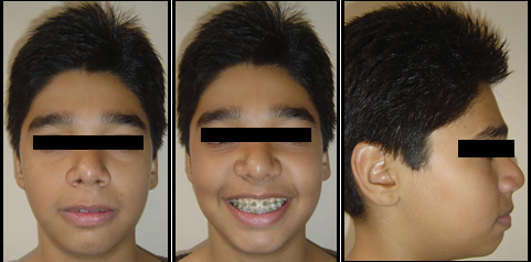
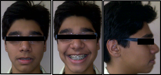
Complete bonding & banding in both maxillary and mandibular arch done, using MBT-0.022X0.028” slot. Initially a 0.012” NiTi wire was used which was followed by 0.014, 0.016”, 0.018”, 0.020” Niti archwires following sequence A of MBT. After 6 months of alignment and leveling NiTi round wires were discontinued. Retraction and closure of spaces was then started by use of 0.019” x 0.025” rectangular NiTi followed by 0.019” x 0.025” rectangular stainless steel wires. Reverse curve of spee in the lower arch and exaggerated curve of spee in the upper arch was incorporated in the heavy archwires to prevent the excessive bite deepening during retraction process and also to maintain the normal overjet and overbite. Anchorage was conserved by light retraction forces constantly monitoring the already well settled molar relation. This is the most important step in an Extraction case wherein anchorage conservation is of utmost importance. Retraction and closure of spaces was done with the help of Elastomeric chains delivering light continuous forces and replaced after every 4 weeks due to force decay and reduction in its activity. Finally light settling elastics were given with rectangular steel wires in lower arch and 0.012” light NiTi wire in upper arch for settling, finishing, detailing and proper intercuspation. The reverse Overjet and overbite was corrected with an ideal occlusion at the end of the fixed apppliance therapy. Also the profile of the patient improved significantly from being convex to now more Orthognathic with a pleasant and consonant smile arc on smiling. Also, the Nasolabial angle improved significantly at the end of treatment
Discussion
Treatment of a moderately crowded Class I malocclusion with extractions of all 1st premolars is challenging. A well chosen individualized treatment plan, undertaken with sound biomechanical principles and appropriate control of orthodontic mechanics to execute the plan is the surest way to achieve predictable results with minimal side effects. Class I malocclusion with Bimaxillary Dentoalveolar protrusion might have any number of a combination of the skeletal and dental component. Hence, identifying and understanding the etiology and expression of Class I malocclusion and identifying differential diagnosis is helpful for its correction. The patient's chief complaint was forwardly placed upper and lower front teeth with excessive show of front teeth. The case was of a clearcut bimaxillary dentoalveolar protrusion with severely proclined upper and lower anterior dentition. The selection of orthodontic fixed appliances is dependent upon several factors which can be categorized into patient factors, such as age and compliance, and clinical factors, such as preference/familiarity and laboratory facilities. The execution of all 1st premolar extraction followed by Fixed appliance therapy appropriately resulted in an improvement in the patient's convex profile in this case. The most important point to be highlighted here is the decision to extract the premolars. After analysing the case thoroughly and reading all pretreatment cephalometric parameters along with evaluating the patients profile clinically, a decision was made of extracting the 1st premolars. Proximal stripping with retraction and closure of spaces could not be executed in this case as this would not adress all the patient problems at the end of the treatment. The patient had excessive proclination of maxillary and mandibular anterior teeth along with crowding in the upper arch. Also the patient had a convex profile with an acute nasolabial angle and a severely decreased Interincisal angle. All these findings made it essentially imperative to extract all 1st premolars. This case could not be managed by non extraction or proximal stripping. Extractions also very efficiently improved the patients profile changing it from being convex to more orthognathic at the end of the treatment. There was improvement in occlusion, smile arc, profile and position of chin. Successful results were obtained after the fixed MBT appliance therapy within a stipulated period of time. The overall treatment time was 16 months. After this active treatment phase, the profile of this 15 year old male patient improved significantly as seen in the post treatment Extra oral photographs. Removable Beggs retainers were then delivered to the patient along with fixed lingual bonded retainers in upper and lower arch.
Table 3
Comparison of pre and post treatment cephalometric readings
Conclusion
This case report shows how Bimaxillary Dentoalveolar Protrusion case can be managed with Extraction of 4 premolars by means of appropriate use of simplified fixed orthodontic treatment and efficient conservation of anvchorage at the same time. The planned goals set in the pretreatment plan were successfully attained. Good intercuspation of the teeth was maintained with class I molar relationship. Treatment of the Prognathic appearing upper and lower jaw included the retraction and retroclination of maxillary and mandibular incisors with a resultant decrease in soft tissue procumbency and facial convexity. The profile changed from convex to orthognathic and the bilateral single tooth crossbite with the maxillary lateral incisors was corrected. The maxillary and mandibular teeth were found to be esthetically satisfactory in the line of occlusion. Patient had improved smile and Profile. The correction of the malocclusion was achieved, with a significant improvement in the patient aesthetics and self-esteem. The patient was very satisfied with the result of the treatment.


