Introduction
To achieve functional efficiency, aesthetic harmony and structural balance is aim of an orthodontic treatment. For ideal result and difficult orthodontic tooth movement 1, 2 anchorage i.e., resistance towards unwanted tooth movement3 is an integral part of an orthodontic treatment.
Bialveolar protrusion is a condition that is mainly characterized through an increased procumbency of lips as well as protrusive and proclined upper and lower anteriors. It is also a usual finding among various different populations.4, 5, 6, 7, 8, 9, 10
Retroclination as well as retraction of maxillary and mandibular incisors with an upshot decrease in soft tissue procumbency and convexity is goal of an orthodontic treatment.11
Regular treatment approach for patients with Class II bialveolar protrusion is extraction of right and left 1st or 2nd maxillary or mandibular bicuspids, followed by retraction of anterior teeth using different anchorage mechanics.12, 13, 14
However, the treatment plan becomes more complex and controversial when a patient has hopeless mandibular first molars that should be extracted and wants to preserve mandibular premolars.
Skeletal anchorage was achieved since TADs were introduced since last two decades.
Present day use of TADs in advanced orthodontics enables an orthodontist to achieve skeletal anchorage that mainly allows them to attain difficult tooth movement.
As a part of TADs miniscrews are being used in various day to day orthodontic practices and it also helps to attain various difficult tooth movement such as intrusion, extrusion, distalization of molars.
When miniscrews were rightfully used various difficult orthodontic cases were reported treated successfully.15, 16, 17, 18, 19
Bialveolar protrusion cases mainly requires levelling as well as alignment of teeth followed by retraction and retroclination of maxillary and mandibular anterior that finally results in a decreased convexity and soft tissue procumbency.
This case report describes orthodontic treatment of an adult female patient with severe bialveolar protrusion along with incompetent lips and compromised mandibular first molars with use of miniscrews (TADs) for anchorage.
Case Report
A 21-year old female patient reported to the clinic with forwardly placed upper and lower anterior teeth.
Extraoral examination revealed, an acute nasolabial angle along with a convex profile, incompetent as well as protrusive upper and lower lips, a positive tongue thrust and mentalis muscle strain.
Intraoral examination revealed that the patient had a poor oral hygiene, with grossly carious right and left mandibular first molars ie, (3-6, 4-6).
All permanent teeth were present through third molar, except for the mandibular right and left first molar which were grossly carious and in a non-restorable status. A Class II canine relationship on the both right and left side was observed.
Angles’s molar relationship on right side could not be determined because of the missing structure of right first mandibular molar due to carries while a class II molar relationship was observed on left side.
In relation with facial midlinethe lower midline was shifted 1 mm to the right. There was also 3-mm crowding in mandibular arch whereas 2mm crowding was found to be in maxillary arch. (Figure 1)
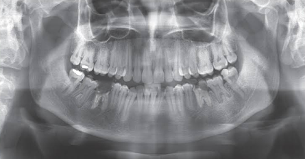
No bone pathology and a normal morphology of condyle was observed in panoramic radiograph. All permanent teeth are present including all four wisdom teeth, there is also presence of severe decay on the mandibular right and left first molar i.e. (3-6, 4-6). (Figure 2).
The lateral cephalogram and its tracing showed dental Class II bialveolar protrusion, but Class I skeletal pattern. The skeletal pattern was normodivergent as evidenced by the FMA (Frankfort mandibular plane angle) of 28º. ANB-2°, SNA-75° and SNB-73° with proclined upper and lower anterior. (Figure 3)
The IMPA (incisor mandibular plane angle) of (100º) reflected the proclination of lower incisors. There were no significant signs or symptoms of temporomandibular disorders. (Figure 3)
Treatment Plan
Extracting mandibular first molars ie, (3-6, 4-6) and maxillary first bicuspids ie, (1-4, 2-4).
Placement of miniscrews during the alignment stage.
En-masse retraction of upper and lower anterior teeth in to the extraction spaces via miniscrews (TADs).
Finishing and detailing finally followed by retention.
Treatment Progress
After extraction of mandibular 1st molars (3-6, 4-6) and maxillary right and left 1st bicuspids, MBT appliance (0.022'' slot) were used.
Levelling and alignment were performed with 0.014'' Niti wires followed by 0.017'' X 0.025'' and finally 0.019'' x 0.025'' Niti wires were used respectively. The patient was given a tongue crib appliance to intercept the tongue thrusting habit. Since the patient was an adult, she was given a removable tongue crib and was also advised to perform tongue exercises like elastic swallow.
Four orthodontic mini-implants of conical shape, 8mm length and 1.4 mm diameter were placed interradicularly between the maxillary second bicuspids and first molars and mesial to mandibular second molar during the alignment stage. (Figure 4).
A 0.017'' × 0.025'' inch Stainless steel arch-wire was placed in upper and lower arches, a closed power chain were applied from the maxillary and mandibular mini-implants to the anterior hook of all four canines to retract the anterior teeth.
After retraction, the treatment was completed with ideal arch wires sequencing and with use of settling elastics (Figure 4). For retention lingual bonded retainers were bonded to the lingual sides of the six anterior teeth. The total treatment time was 22 months.
Angles Class I canine relationship was achieved bilaterally with well-coordinated upper and lower arches. Significant reduction in dentoalveolar protrusion was seen due to retraction of upper and lower anterior completely into the extraction spaces (Figure 5).
Superimposition of the pre and post treatment cephalometric analysis showed that both maxillary and mandibular incisors were bodily retracted (U1 to NA 24°/ 5mm, L1 to NB 28° / 4.5mm). Also, there was no significant change in ANB angle. Esthetic line in upper lip changes from 1 mm to -2 mm while a considerable 2mm to -1 mm in lower lip. No considerable change in Frankfurt mandibular plane angle from previous 28° to post 27° was observed suggesting normal direction of lower facial growth both horizontally and vertically, improvement in incisor mandibular plane angle was observed from previous 100º to 96º post treatment.
Effective retraction of Upper and lower lip was achieved that finally lead to increase in nasolabial angle from 85° to 97° resulted in a significant profile change of patient.
Table 1
Cephalometricparameters
Discussion
Patients consider for orthodontic treatment reason being facial aesthetics especially in bialveolar protrusion cases that are mainly recognized by procumbency of upper and lower lips along with flaring of the maxillary and mandibular anterior.20
In orthodontics, extracting first permanent molars to resolve bialveolar protrusion cases is not widely accepted.21
Mills study indicated that extracting first molars could reduce the prognosis by half and doubles the treatment time.21
However, in this case, extraction of mandibular right and left first permanent molars were performed because of their non restorable status caused by extensive carries and that finally indicated for extraction.
Other treatment options included such as extracting the lower two premolars i.e, (3-4, 4-4) but that could not possible due to grossly carious mandibular right and left first molars i.e, (3-6, 4-6), thus having premolars against molars might not be a favourable treatment of choice.
Many clinical necessities let unusual extraction of molar including extensive caries and large restorations.
Molar extraction is usually indicated when
Severe carious lesions, ectopic eruption, or is considered in severe rotation.
Facial profiles with Moderate arch length deficiencies.
Distalization of molars in relapsed orthodontic cases for space gaining. 22
Removing molars can serve as an alternative for extraction of the maxillary or mandibular bicuspids.
The clinical efficacy23, 24 as well as stability25 of temporary skeletal anchorage devices (TADs) have been widely described and it is considered as a highly efficient method for solving various orthodontic problems and difficult tooth movements that cannot be corrected using conventional orthodontic methods. Several skeletal anchorage devices that are efficient in controlling anchorage have been developed to obtain anchorage control during the distalization movement. Various Temporary Anchorage Devices (TADs) such as Endosseous implants,26 Surgical miniplates,27 onimplants,28 palatal implants,29 surgical miniscrews,30 helped to overcome various limitation of difficult orthodontic tooth movements.
Miniscrews (TADs) can provide anchorage in missing or compromised molar cases mainly to facilitate the retraction of anterior in to extraction spaces.
High possibility and risk factor involved in direct anchorage as a miniscrew could fail during en-masse retraction and in the presence of power chain, the canine might be pushed mesially. Thus for this purpose a close follow-up was an important aspect of an orthodontic treatment, specifically during the first 2 weeks of retraction.
For En-masse retraction of anterior teeth skeletal anchorage with use of miniscrews was important to implement this treatment plan.
Current study enabled us to effectively retract upper and lower anterior and eventually upper and lower lips to a more favourable position when anchorage was properly maintained with the use of miniscrews (TADs).
Thus the use of miniscrews (TADS) facilitated the treatment of Class II bialveolar protrusion with compromised mandibular molar cases more effectively regardless the extraction pattern used.
Conclusion
The total treatment period was 22 months. Use of miniscrews (TADs) in Class II bialveolar protrusion cases with compromised molars can help to achieve and provide skeletal anchorage for en-masse retraction of the anterior teeth into the extraction spaces resulting in reduction in procumbency of lips and retroclination of upper and lower anterior that resulted in a significant profile change of patient.


