- Visibility 402 Views
- Downloads 45 Downloads
- Permissions
- DOI 10.18231/j.ijodr.2024.043
-
CrossMark

Evaluation of width of buccal corridor space in skeletal class I, class II, and class III malocclusion: A photographic study
Abstract
Aim & Objectives: To evaluate width of buccal corridor space in patients with skeletal class I, class II and class III malocclusion and to find out whether the skeletal malocclusion in sagittal plane can affect the width of buccal corridor space.
Introduction: Smile analysis has been treated as a separate entity from cephalometrics and cast analysis in orthodontic diagnosis and treatment planning. The transverse dimension of smile refers to the broadness of smile or the presence or absence of buccal corridor space. Buccal corridor space is nothing but the distance between the distal point of the canines and the lateral junction of upper and lower lips during smiling. Buccal corridor space has been thought of primarily in terms of maxillary width, but there is also evidence that they are heavily influenced by the anteroposterior position of maxilla in relation to lip drape.
Materials and Methods: 30 patients between the ages of 18 to 30 years were included in the study. Based on ANB angle this patient were divided into 3 groups i.e., Skeletal class I, class II and class III malocclusion. Photographic analysis:Posed smile photographs of patients of age between 18 to 30 years made under standardized conditions were used for analysis to determine buccal corridor space.
Results: Transverse dimension is said to be a function of both arch width and antero-posterior position of the maxillary and mandibular arches.The results of this study show that there exist a correlation between malocclusion in sagittal plane and the amount of buccal corridor space. This fact must be considered during the orthodontic evaluation, diagnosis and planning of treatment for individuals with different types of malocclusions.
Conclusion: This study's results show a correlation between malocclusion in sagittal plane and the amount of buccal corridor space. This fact must be considered during the orthodontic evaluation, diagnosis, and planning of treatment for individuals with different types of malocclusions.
Introduction
Face is one of the key features of physical attractiveness of an individual. Studies have reported face as being the most important component of aesthetic perception of any person. Mouth and eyes are most important components for determining aesthetic value in the face. Since aesthetic consideration is one of the foremost reasons for patients to undergo orthodontic treatment, it becomes the job of orthodontist to properly evaluate and understand the factors influencing aesthetics of any person.
The smile aesthetics is one of the most important contributors to the facial aesthetics. Further the factors contributing to smile aesthetics include the area of gingival display; colour, contour, texture and height of the gingiva; the presence of smile arc; the teeth by its contributing factors of size, shape, shade and alignment and the buccal corridor space.
Despite the fact that there have been many soft tissue evaluations of the face, the majority of them.[1], [2], [3], [4], [5], [6], [7], [8] focused on the sagittal plane's soft tissue profile. Nonetheless, the significance of the aesthetics in the frontal view was highlighted by Proffit. [9] Arnett and Bergman. [7] Arnett et al., and Arnett et al. As a result, orthodontists must shift their focus during patient evaluation from the sagittal plane to the frontal plane while planning and assessing orthodontic therapy. [10] Orthodontic patients also worry about how they seem both dynamically when they smile and converse, in addition to how they look static. [11], [12], [13], [14], [15]
It is important to differentiate between the social smile and the enjoyment smile. The social smile is a voluntary smile a person uses in social settings or when posing for a photograph. When you are introduced to someone, your smile indicates that you are friendly and “pleased to meet” that person. The enjoyment smile is an involuntary smile and represents the emotion you are experiencing at that moment. The enjoyment smile therefore has many descriptors, such as laughing, wry, knowing, or insipid.In orthodontic treatment, the socially posed smile is known as "smile designing" since it is reproducible and repeatable. [12], [16], [17], [18], [19] One of the evaluating points for smile aesthetics is the buccal corridor. It is a dark or black area that is visible when smiling and is located between the maxillary lateral teeth and the corner of the mouth.
Evaluation of buccal corridor width in patients with skeletal class I, class II, and class III malocclusion was the goal of this study.
Materials and Methods
Thirty patients with skeletal malocclusion in the sagittal plane, aged 18 to 30, were selected from the Department of Orthodontics and Dentofacial Orthopedics. They were male and female.
Inclusion criteria
Individuals in the age group of 18-30 years of any gender.
Individuals with Skeletal malocclusion in Sagittal plane.
No/ minimal crowding, spaces or rotations.
Exclusion criteria
Previous history of orthodontic treatment.
Individuals with craniofacial anomaly.
Individuals with crossbite and scissor bite.
An enormous prosthesis or missing teeth.
Cephalometric analysis
Lateral cephalograms were made with Frankfort horizontal plane maintaining parallel to the floor. Acetate paper measuring 0.003 inches was used to trace the lateral cephalograms.
These patients were categorized into three groups, skeletal class I, class II, and class III malocclusion, based on the ANB angle.
Photographic analysis
The buccal corridor space was determined by analyzing pictures of patients' posed smiles that were taken under standardized conditions. The patients were between the ages of 18 to 30.
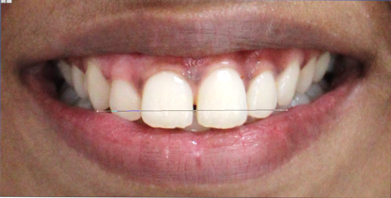
The measurements of the inter canine width([Figure 1]) and inter commissural width([Figure 2]) were made in 0.01 mm units using Adobe Photoshop's linear measuring tool.
The formula below was used to determine the buccal corridor linear ratio(BCLR) using the linear measurement tool in Adobe Photoshop 7.0 software:
BCLR=Upper inter canine width Inter commissural width×100
Group 1: BCLR in patients with skeletal class I malocclusion.
Group 2: BCLR in patients with skeletal class II malocclusion.
Group 3: BCLR in patients with skeletal class III malocclusion.
The buccal corridor linear ratio(BCLR)([Figure 3]) in skeletal class I, II, and III malocclusion were compared.

Results


|
|
|||
|
|
N |
Mean |
Std. Deviation |
|
Group I |
10 |
57% |
0.01 |
|
Group II |
10 |
56.10% |
0.03 |
|
Group III |
10 |
56.50% |
0.03 |
Discussion
When compared to the Class I group, it was discovered that buccal corridor space was greater in Class II subjects([Figure 4]). This might be as a result of the narrower maxillary arch of Class II Division 1 participants compared to Class I participants.
A larger buccal corridor space is usually the consequence of a narrower arch. This was consistent with earlier research by Ackerman et al., Sarver and Ackerman, Snyder.[20] McNamara, and Ackerman et al.[21]
The greater portion of the dental arch is positioned more posteriorly with respect to the anterior oral commissure in cases of skeletal class III malocclusion, in which the maxilla is retrusive.
This creates the impression of a wider buccal corridor in the frontal dimension. When this occurs, there is less "negative space" because a larger section of the maxilla is positioned into the buccal corridor as a result of maxillary advancement.
Buccal corridor space is affected by various hard and soft tissue factors. A study by Ackerman et al noted that the antero-posterior position of the maxilla and the rotation of the upper first molars could be the influencing factors on buccal corridor area. Snyder, Sarver and Ackerman noted that the wider the inter canine and inter premolar width, the smaller the buccal corridor space. Study conducted by Yang et al., [22] showed that long face individuals with high Mandibular plane angle and increased facial height tend to have less buccal corridor space.
According to Sarver and Ackerman, [13], [15] the facial types near two ends of the spectrum, dolichocephaly and brachycephaly, are greatly affected by a positional change in tooth mass in the buccal corridor. According to Yang et al.[22] to control the amount of buccal corridor area for achieving a better esthetic smile, it is necessary to observe the vertical pattern of the face, amount of upper incisor exposure, and sum of the tooth material.
Conclusion
The sagittal position of the maxillary and mandibular arches as well as their width are considered to influence transverse dimension.The results of the study show a correlation between malocclusion in the sagittal plane and the amount of buccal corridor space.It is important to consider this information when diagnosing, treating, and evaluating patients with various malocclusions in orthodontics.
Limitations
One of the study's limitations is that it only included a limited number of samples. Our understanding of the width of buccal corridor space in various sagittal malocclusions will improve with more research with a greater number of samples.
Ethical Approval
4090/2023 dated 12/10/23
Source of funding
None.
Conflict of Interest
None.
References
- Burstone C. The integumental profile. Am J Orthod. 1958;44(1):1-25. [Google Scholar]
- Ricketts R. Cephalometric synthesis. Am J Orthod. 1960;46(9):647-73. [Google Scholar]
- Merrifield L. The profile line as an aid in critically evaluating facial esthetics. Am J Orthod. 1966;52(11):804-26. [Google Scholar]
- Scheideman G, Bell W, Legan H, Finn R, Reisch J. Cephalometric analysis of dentofacial normals. Am J Orthod. 1980;78(4):404-20. [Google Scholar]
- Holdaway R. A soft-tissue cephalometric analysis and its use in orthodontic treatment planning. Part I. Am J Orthod. 1983;84(1):1-28. [Google Scholar]
- Powell N, Humphreys B. . Proportions of the Aesthetic Face. 1984. [Google Scholar]
- Arnett G, Bergman R. Facial keys to orthodontic diagnosis and treatment planning. Part I. Am J Orthod Dentofacial Orthop. 1993;103(4):299-312. [Google Scholar]
- Arnett G, Jelic J, Kim J, Cummings D, Beress A. Soft tissue cephalometric analysis: diagnosis and treatment planning of dentofacial deformity. Am J Orthod Dentofacial Orthop. 1999;116(3):239-53. [Google Scholar]
- Proffit W. The soft tissue paradigm in orthodontic diagnosis and treatment planning: a new view for a new century. J Esthet Dent. 2000;12(1):46-9. [Google Scholar]
- Kerns L, Silveira A, Kerns D, Regennitter F. Esthetic preference of the frontal and profile views of the same smile. J Esthet Dent. 1997;9(2):76-85. [Google Scholar]
- Ackerman J, Proffit W, Sarver D. The emerging soft tissue paradigm in orthodontic diagnosis and treatment planning. Clin Orthod Res. 1999;2(2):49-52. [Google Scholar]
- Ackerman M, Ackerman J. Smile analysis and design in the digital era. J Clin Orthod. 2002;36(4):221-36. [Google Scholar]
- Sarver D, Ackerman M. Dynamic smile visualization and quantification: part 1. Evolution of the concept and dynamic records for smile capture. Am J Orthod Dentofacial Orthop. 2003;124(1):4-12. [Google Scholar]
- Sarver D, Ackerman M. Dynamic smile visualization and quantification: part 2. Smile analysis and treatment strategies. Am J Orthod Dentofacial Orthop. 2003;124(2):116-27. [Google Scholar]
- Ackerman M, Brensinger C, Landis J. An evaluation of dynamic lip-tooth characteristics during speech and smile in adolescents. Angle Orthod. 2004;74(1):43-50. [Google Scholar]
- Hulsey C. An esthetic evaluation of lip-teeth relationships present in the smile. Am J Orthod. 1970;57(2):132-44. [Google Scholar]
- 3rd OR, Sperry T, Begole E. The influence of facial animation in smile characteristics. Int J Adult Orthodon Orthognath Surg. 1988;3(4):233-9. [Google Scholar]
- Morley J. Smile design terminology. Dent Today. 1996;15(6). [Google Scholar]
- Ackerman J, Ackerman M, Brensinger C, Landis J. A morphometric analysis of the posed smile. Clin Orthod Res. 1998;1(1):2-11. [Google Scholar]
- Snyder R. Class II malocclusion correction: An American board of orthodontics case. Am J Orthod Dentofacial Orthop. 1999;116(4):424-9. [Google Scholar]
- Mcnamara J. Maxillary transverse deficiency. Am J Orthod Dentofacial Orthop. 2000;117(5):567-70. [Google Scholar]
- Yang I, Nahm D, Baek S. Which hard and soft tissue factors relate with the amount of buccal corridor space during smiling?. Angle Orthod. 2008;78(1):5-11. [Google Scholar]